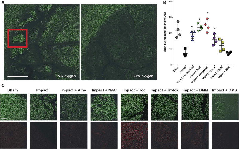
Fig. 5. Acute loss of mitochondrial content after impact is prevented by inhibition of electron transport or critical redox events.
(A) Representative confocal micrographs at 4X magnification showing the site of a 2 J impact to bovine femoral osteochondral explants 24 hours after injury in either 5 or 21% oxygen. Scale bar, 1 mm. Red inset square indicates zone of interest for quantitation (B) and images shown in (C). (B) Quantitation of MitoTracker Deep Red staining 24 hours after 2 J impact (n = 4; * P < 0.01 versus impact alone via one-way ANOVA). Data represent the mean with SD shown. AU, arbitrary units. (C) Representative 10X micrographs of areas within the impact site as indicated by the red square in (A) and an analogous region central to the sham (unimpacted) osteochondral explant was chosen. Scale bar, 100 µm. Calcein AM is shown in green in the top row and MitoTracker Deep Red is shown in red in the bottom row. Amobarbital, NAC, α-tocopherol (Toc), trolox, dimethylmalonate (DMM), and dimethyl succinate (DMS) were all tested. Dark areas within the images indicate cracks or other structural damage from the impacts.

