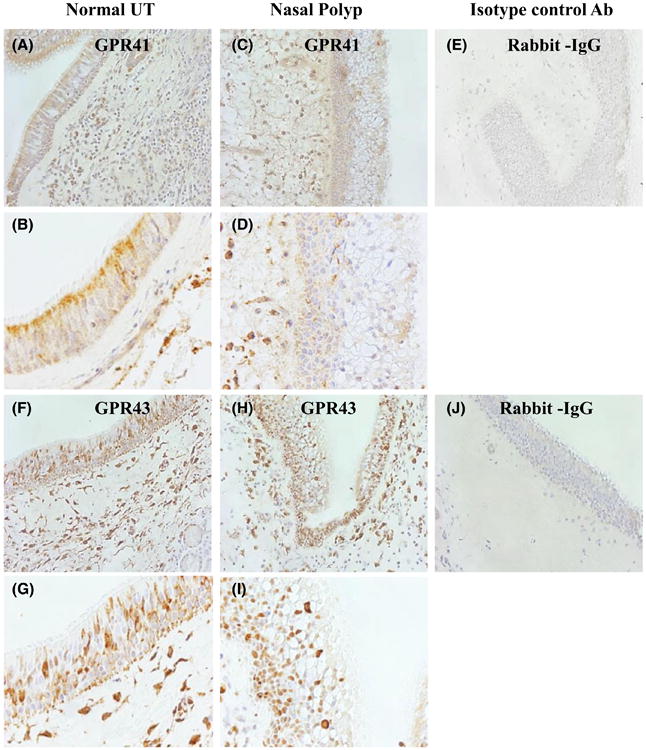
Figure 1.

Immunohistochemical (IHC) detection of GPR41 (A-E) and GPR43 (F-J) in human sinonasal tissue. Representative immunostaining for GPR41 in UT from control subject (A, B) and in NP tissue (C, D), and staining for GPR43 in UT from a control subject (F, G) and in NP tissue (H, I). Representative isotype control antibody staining in NP tissue is shown (E, J). Magnification ×200 (A, C, E, F, H, J) and ×400 (B, D, G, I)
