Fig. 1.
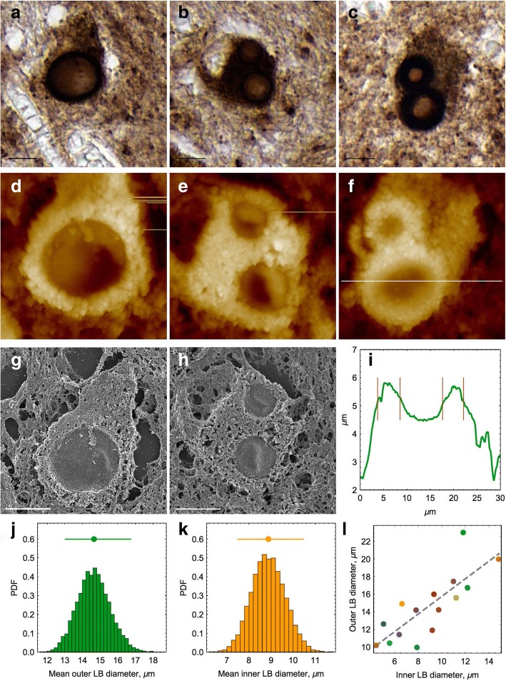
Microscopy of α-syn Lewy bodies in the PD substantia nigra (an individual case). a–c Representative intracellular Lewy bodies immunostained with α-syn antibodies. Lewy bodies are shown in dark brown color and the host cells in lighter brown shade. Scale bars equal to 10 μm. d–f AFM height images of the corresponding Lewy bodies (from a–c). The surfaces of Lewy bodies and surrounding tissues are covered with DAB crystals used in immunohistochemical procedure to stain the tissue samples (shown in light color). Image sizes are 20 × 20 μm. g, h Scanning electron microscopy images of Lewy bodies shown in a, b. i AFM cross-section of Lewy body; its position is shown in f by white line. j, k Distribution of Lewy body mean outer and inner diameters, respectively, calculated by using BCa technique from AFM data. Mean diameters and their 95% CI are shown above the histograms. Probability density function (PDF) is shown along the y-axis. l Linear dependence between the inner and outer diameters of the Lewy bodies analyzed by AFM. Each point represents individual randomly selected Lewy body from the same patient and is shown in individual color

