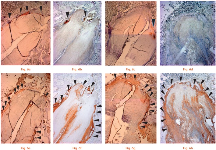
Photomicrographs of the tendon/bone interface with immunohistochemical staining for cartilage (type II collagen) (a-d) (magnification × 25). Cartilage tissue (arrowheads) was observed at four weeks in a) the Socket group and b) Tunnel group. At eight weeks, staining for cartilage was reduced in both the c) Socket group and d) Tunnel group. e-h) Photomicrographs of the tendon/bone interface with immunohistochemical staining for type III collagen (magnification × 25). Type III collagen (equivalent to Sharpey’s fibres; arrowheads) was primarily expressed at the tendon/bone interface at four weeks in the e) Socket and f) Tunnel groups. Staining was similar at eight weeks in the g) Socket and h) Tunnel groups (scale bars, 1.0 mm).

An official website of the United States government
Here's how you know
Official websites use .gov
A
.gov website belongs to an official
government organization in the United States.
Secure .gov websites use HTTPS
A lock (
) or https:// means you've safely
connected to the .gov website. Share sensitive
information only on official, secure websites.
