Figure 6.
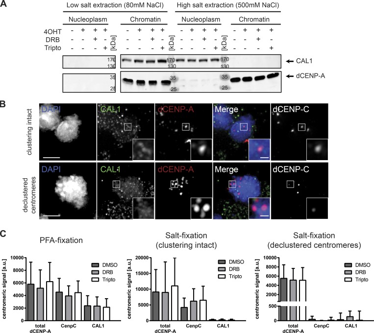
Centromere association of CAL1 is less stable than CENP-C, and neither CAL1 nor CENP-C is sensitive to transcriptional inhibition. (A) Western blot analysis showing the displacement of CAL1 from chromatin after high-salt extraction. Arrows mark protein of interest. Endogenous dCENP-A serves as a marker for chromatin. (B) Maximum-intensity projection of cells fixed after 30 min of 0.5 M salt extraction and immunostained for CAL1 and dCENP-C. Extracted cells with clustering of centromeres still intact (upper panel) and disrupted (lower panel) are shown. Bars, 3 µm. (C) Quantifications of centromeric localization of total dCENP-A, dCENP-C, and CAL1 in inhibitor-treated cells. Fixation type and group of analyzed cells is indicated above. n = 3 replicates; n = 25–50 cells. Data are mean + SD.

