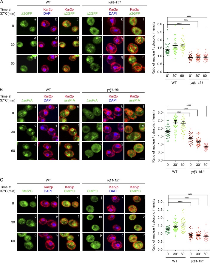
Figure 2.
Localization of misfolded cytosolic proteins is defective in ydj1-151 cells. (A–C) WT and ydj1-151 cells expressing ΔssPrA, Δ2GFP, and Ste6*C were grown to log phase at room temperature and incubated at 37°C for 30 or 60 min. Cells were prepared for indirect immunofluorescence as described in Materials and methods. Substrates were detected using anti-HA antibodies in the green channel. ER and nuclear envelope were visualized in the red channel using anti-Kar2 antiserum. Nuclei were localized using DAPI staining. Cell imaging and acquisition were performed by confocal microscopy. Quantification of fluorescence intensity was done as described in Materials and methods. One-way ANOVA was used to test for significance (35 < n < 50; ****, P < 0.0001). The results shown are representative of two independent experiments. Bars, 2 µm.

