Figure 5.
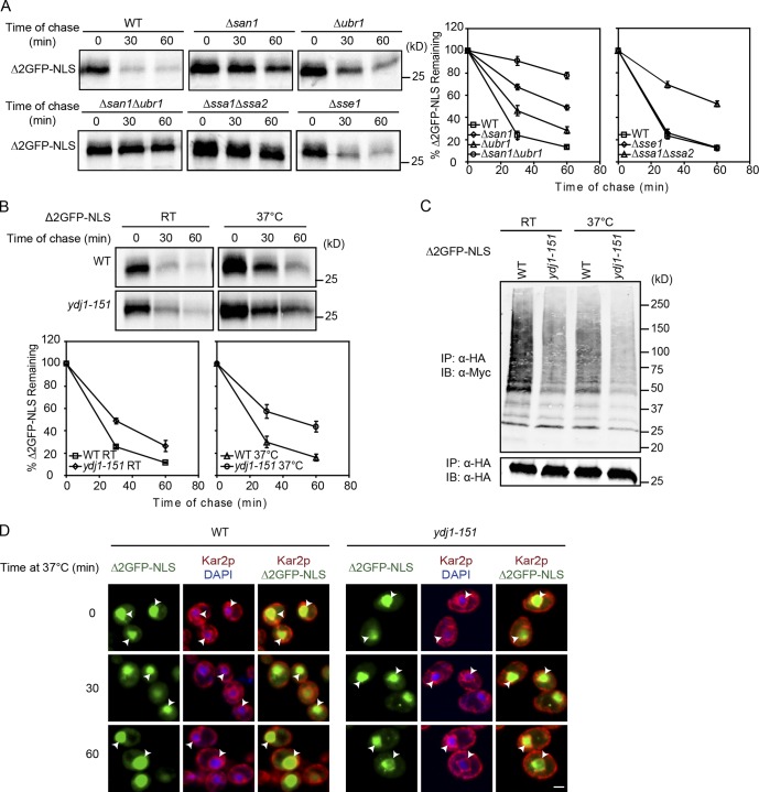
Appending an NLS to Δ2GFP bypasses requirement for Ydj1 and Sse1. (A and B) Turnover of Δ2GFP-NLS in WT, Δsan1, Δubr1, Δsan1Δubr1, Δssa1Δssa2, Δsse1, and ydj1-151 cells was determined by pulse chase at temperatures indicated. All data plotted were processed using Excel, reflecting three independent experiments with the means and SD indicated. (C) Ubiquitination of Δ2GFP-NLS was determined as described in Fig. 1 B. IB, immunoblot; IP, immunoprecipitation. (D) Cells expressing Δ2GFP-NLS were grown to log phase at room temperature and shifted to 37°C for 30 or 60 min. After fixation, Δ2GFP-NLS and Kar2 were visualized by immunostaining with respective primary and secondary antibodies. Nuclei were marked by DAPI staining. Arrowheads indicate positions of nuclei. Bars, 2 µm.

