Figure 5.
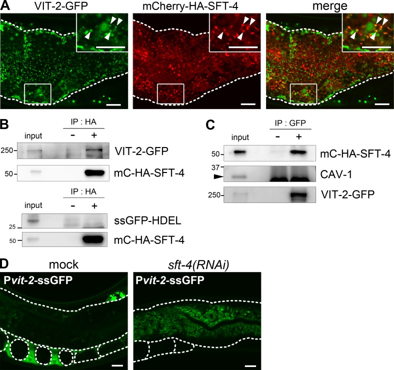
SFT-4 interacts with VIT-2 in vivo. (A) Subcellular localization of VIT-2 and SFT-4 in intestinal cells. VIT-2–GFP partially colocalized with mC–HA–SFT-4 on punctate structures (arrowheads). (B and C) SFT-4 interacts with VIT-2. Lysates of whole animals coexpressing VIT-2–GFP and mC–HA–SFT-4 were immunoprecipitated with anti-HA (B; upper panel) and anti-GFP (C) antibodies. The precipitates and 0.3% of the total lysate were immunoblotted with anti-HA and anti-GFP antibodies. SFT-4 does not interact with ssGFP-HDEL when lysates of whole animals coexpressing ssGFP-HDEL and mC–HA–SFT-4 were immunoprecipitated with anti-HA antibody (B; lower panel). CAV-1 (C; arrowheads) was not coimmunoprecipitated with VIT-2–GFP. (D) Secretion of ssGFP from the intestine into the body cavity was impaired in sft-4(RNAi) animals. Dotted lines indicate the outlines of intestines, oocytes, and embryos. Regions surrounded by squares are enlarged (4×) in insets. Bars, 10 µm.

