Figure 6.
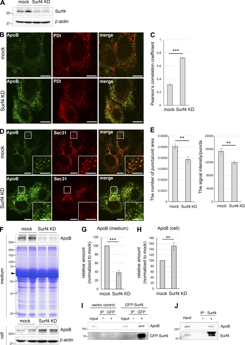
Surf4, a mammalian homologue of SFT-4, is required for ER export of ApoB in HepG2 cells. (A) Whole-cell lysates were immunoblotted with antibodies against Surf4 and β-actin. Surf4 expression was efficiently silenced. (B) Surf4 loss causes ApoB accumulation in the ER. HepG2 cells were transiently transfected for 3 d with control (mock) or surf4 siRNA (Surf4 KD) and then fixed and stained with anti-ApoB and anti-PDI antibodies. In Surf4 KD cells, ApoB was accumulated in the ER and was colocalized with PDI. Bars, 10 µm. (C) Colocalization quantifications were calculated by using Pearson’s correlation coefficient and statistically analyzed using Student’s t test; ***, P < 0.001; error bars: SEM (n = 96 and 87 of mock and Surf4 KD cells, respectively). (D) Loss of Surf4 reduced the number and size of Sec31-positive ERES. HepG2 cells were transiently transfected for 3 d with control (mock) or surf4 siRNA (Surf4 KD) and then fixed and stained with anti-ApoB and anti-Sec31 antibodies. Regions surrounded by squares are enlarged (9×) in insets. Bars: 10 µm; (insets) 5 µm. (E) The quantifications of the number and signal intensity of Sec31-positive punctate structures spread in the cytoplasm were measured and statistically analyzed using Student’s t test; **, P < 0.05; error bars: SEM (left panel, n = 70 and 50 of mock and Surf4 KD cells for the number of puncta/unit area; right panel, n = 62 and 66 of mock and Surf4 KD cells for the signal intensity/puncta, respectively). (F) The pattern of total secreted proteins. Whole-cell lysates and culture medium were immunoblotted with anti-ApoB. Coomassie Brilliant Blue staining of culture medium is also shown in middle panel. The secretion was not generally inhibited by the loss of Surf4. The arrowhead presumably indicates BSA conjugated with oleic acids. (G and H) ApoB amount was quantified through densitometric scanning of band intensities, and the relative amount was determined. The amount of ApoB secreted from Surf4-depleted cells was significantly decreased as compared with that secreted from mock-treated cells (G), whereas the amount of ApoB in whole cell lysates was significantly increased (H). Results were analyzed using Student’s t test; **, P < 0.05; ***, P < 0.001; error bars: SEM, n = 6 (G) or 4 (H) are shown. (I) Lysates from vector control or GFP-Surf4 transfected HepG2 cells were immunoprecipitated with anti-GFP antibody. The precipitates and 1% of the total lysate were immunoblotted with anti-ApoB antibody. (J) HepG2 cell lysates were immunoprecipitated with anti-Surf4 antibody. The precipitates and 1% of the total lysate were immunoblotted with anti-ApoB antibody.

