Figure 4.
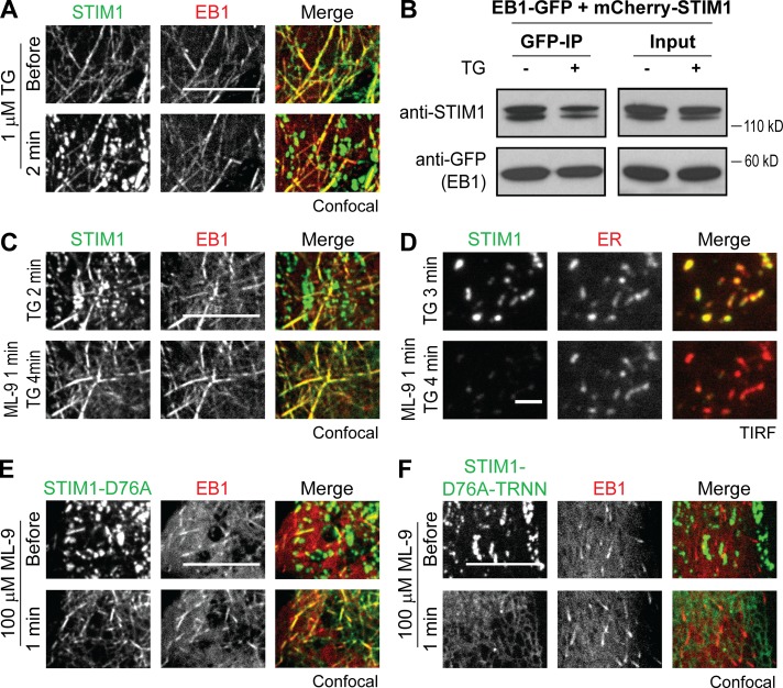
Activated STIM1 retains EB1 binding ability in ER Ca2+-depleted cells. (A) Localization of YFP-STIM1 and EB1-mCherry in HeLa cells during the resting state and after 1 µM TG treatment, monitored by confocal microscopy. (B) IP of EB1-GFP with mCherry-STIM1 after 1 µM TG treatment in HeLa cells. Protein levels of EB1-GFP and mCherry-STIM1 in total cell lysates (Input) and IP were assessed by Western blotting using antibodies against GFP and STIM1. (C) Colocalization of YFP-STIM1 and EB1-mCherry in HeLa cells after 100 µM ML-9 treatment during ER Ca2+ depletion by 1 µM TG, monitored by confocal microscopy. (D) Disruption of TG-induced YFP-STIM1 accumulation at ER–PM junctions labeled by mCherry-ER in HeLa cells after 100 µM ML-9 treatment, monitored by TIRF microscopy. (E) Colocalization of YFP-STIM1-D76A and EB1-mCherry in HeLa cells after 100 µM ML-9 treatment, monitored by confocal microscopy. (F) YFP-STIM1-D76A-TRNN displayed ER localization without colocalizing with EB1-mCherry in HeLa cells after 100 µM ML-9 treatment, monitored by confocal microscopy. Bars: (A, C, E, and F) 10 µm; (D) 2 µm.

