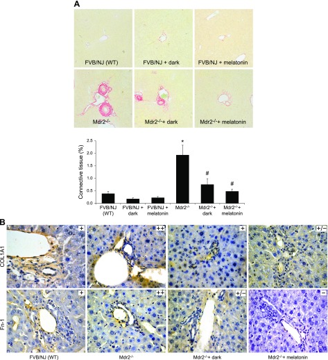
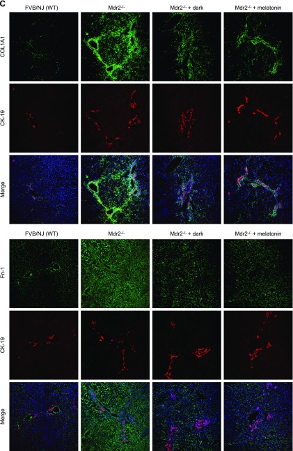
Figure 3.
A–C) In Mdr2−/− mice, fibrosis was enhanced (A) and immunoreactivity was increased for COL1A1 and Fn-1 (B, C), compared with WT mice in which they were reduced by exposure to the dark or melatonin (10 different fields analyzed from each sample from 3 different animals). Dark exposure or melatonin treatment did not alter fibrosis in normal WT mice (A). Original magnification, ×25. D) Expression of Col1a1, Fn-1 and Tgf-β1 was increased in total liver and cholangiocytes and stellate cells from Mdr2−/− mice compared with WT mice, and was reduced by exposure to the dark or melatonin. Data are means ± sem of 6 evaluations in total liver samples collected from 6 separate animals, in cholangiocytes collected from 3 cumulative preparations from 4 mice (n = 12 mice), and in LCD-isolated stellate cells from 3 individual mice. *P < 0.05 vs. WT mice, #P < 0.05 vs. Mdr2−/− mice.


