
Figure 2.
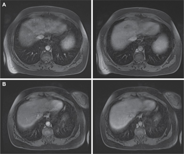
Interval MRI images from Case 1.
Notes: (A) Early arterial and late portal venous phase fat-saturated postcontrast T1 imaging demonstrating an early enhancing intrahepatic mass with washout involving segments IV, VII, and V consistent with diffuse HCC obtained at diagnosis. (B) Repeat early arterial and late portal venous phase fat-saturated postcontrast T1 imaging obtained 12 weeks later with no subsequent HCC therapy demonstrating substantially decreased tumor burden.
Abbreviation: HCC, hepatocellular carcinoma.