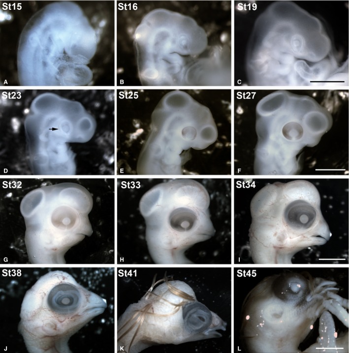
Figure 1.

Stereomicroscope images of some of the embryos included in the present study showing the external gross anatomical changes of the eye. The Taeniopygia guttata embryos were staged according developmental stages (St) established by Murray et al. ( 2013 ). The optic cup was clearly distinguishable between St15 and St23 (A–D). A faint pigmentation in the RPE was first observed at St23 in the caudal region of the optic cup (arrow in D). At St27, the eye was completely pigmented (F). From St38 until perinatal stages, the eyelids progressively covered the eye (J,K,L). RPE, retinal pigment epithelium. Scale bars: 2 mm (A–D); 4 mm (E,F); 6 mm (G); 7 mm (H); 10 mm (I–K).
