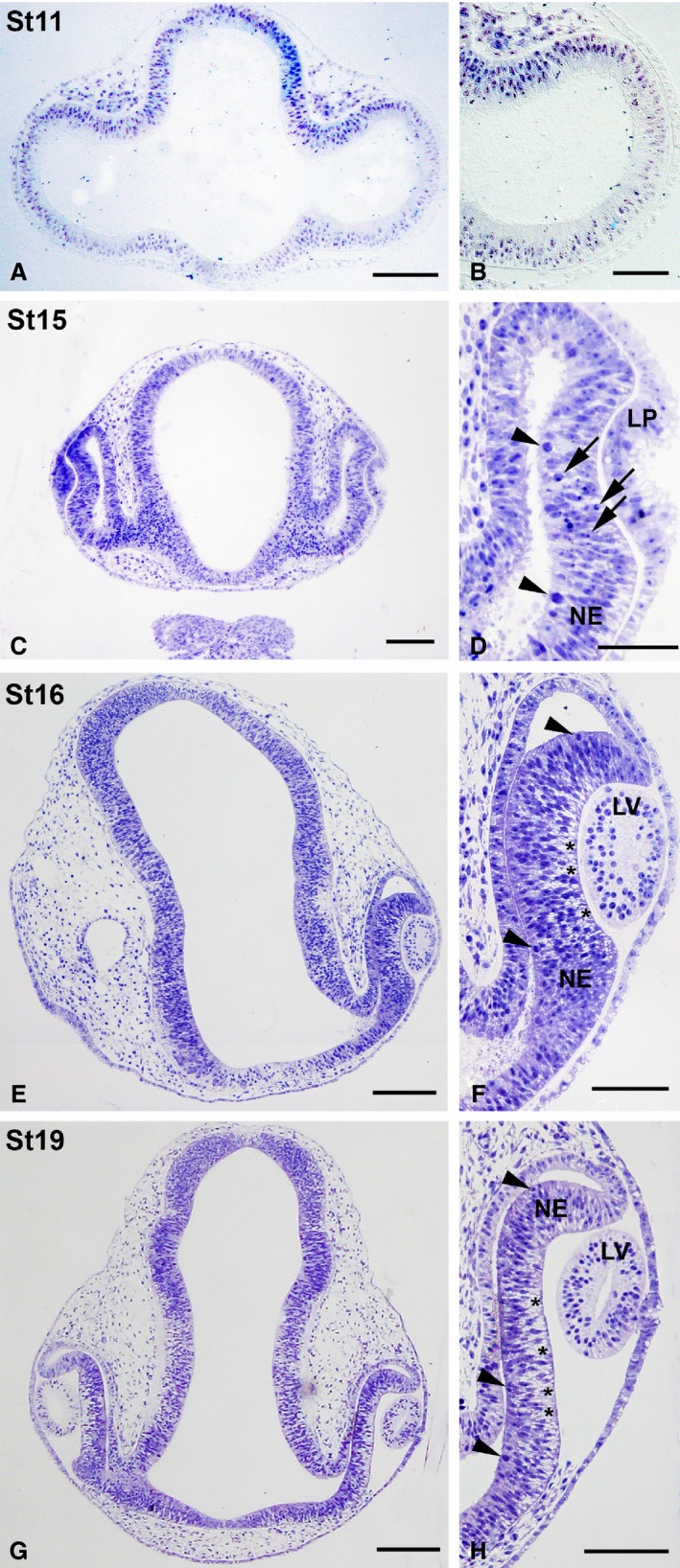
Figure 5.

Toluidine blue‐stained semi‐thin sections showing early stages of development of the eye rudiment in Taeniopygia guttata. Optic vesicles were composed of an undifferentiated neuroepithelium at St11 (A, B). Progressive invagination of the distal optic vesicle resulted in the formation of the optic cup (C–H). Many mitotic figures were seen in the scleral surface of the presumptive neural retina by these stages (arrowheads in D,F,H). The lens placode was present at St15 (C,D) and the lens vesicle could be observed by St16–St17 (E–H). Abundant pyknotic bodies (arrows in D) were observed in the presumptive neural retina at St15 (C,D) and extracellular spaces near the vitreal surface were clearly distinguishable by St16–St17 (asterisks in F,H). LP, lens placode; LV, lens vesicle; NE, neuroepithelium. Scale bars: 100 μm (A–H).
