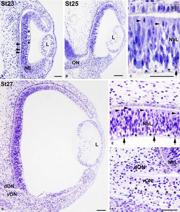
Figure 6.

Toluidine blue‐stained transverse sections of the undifferentiated Taeniopygia guttata retina. Magnification of (B) is shown in (C), and magnifications of (D) are shown in (E,F). Lamination was absent in the developing retina between St23 and St27 (A–E). Abundant ventricular mitosis (arrowheads in A,C,E) and extracellular spaces located near the vitreal surface (asterisks in A,C,E) were observed. The neural retina is composed of NE at St23 and the first differentiated neuroblasts were observed in the vitreal region of the retina at St25 (arrow in C), increasing in number at St27 (arrows in E). Ganglion cell axons were detected in the ventral region of the ON (asterisks in F). Note that faint pigmentation was first observed in the PE at St25. dON, dorsal optic nerve; L, lens; NbL, neuroblastic layer; NE, neuroepithelium; PE, pigment epithelium; vON, ventral optic nerve. Scale bars: 100 μm (A–F).
