Figure 8.

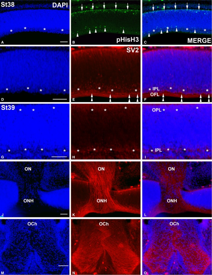
pHisH3‐ (A–C) and SV2‐immunoreactivities (D–O) in the Taeniopygia guttata retina and optic pathways at St38 (A–F) and St39 (G–O). Sections were counterstained with DAPI. pHisH3‐immunopositive mitoses were detected in both the scleral (arrows in B,C) and vitreal regions (arrowheads in B,C). At St38, the IPL could be recognized as a DAPI‐negative layer devoid of cell nuclei in the central retina (asterisks in A,C,D,F) that was faintly labelled with anti‐SV2 antibody (asterisks in E,F). SV2‐immunoreactivity was also detected in sparse ganglion cell perikarya (double arrowheads in E,F) and in the optic fibre layer. A poorly developed SV2‐immunoreactive OPL emerged at St39 in the outer retina, whereas the IPL increased in size (asterisks in G–I). Ganglion cell axons were strongly labelled with the SV2 antisera in both the optic nerve (G–I) and optic chiasm (J–L). IPL, inner plexiform layer; OCh, optic chiasm; OFL, optic fibre layer; ON, optic nerve; ONH, optic nerve head; OPL, outer plexiform layer. Scale bars: 100 μm (A–O).
