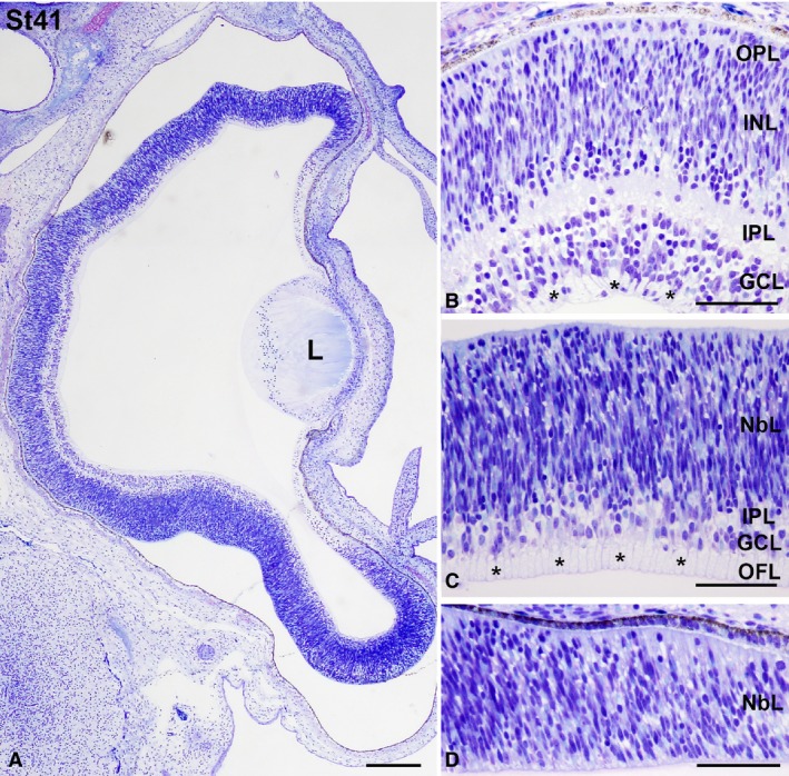
Figure 9.

Toluidine blue‐stained transverse sections at St41 Taeniopygia guttata retina. Magnifications of the central (B), mid‐peripheral (C), and peripheral (D) retina are shown. Abundant ganglion cell axons were observed in the OFL (asterisks in B,C). While retinal lamination was complete in the central retina (A,B), the OPL was absent in the mid‐peripheral retina (A,C). The peripheral retina showed an undifferentiated aspect (A, D). GCL, ganglion cell layer; INL, inner nuclear layer; IPL, inner plexiform layer; L, lens; NbL, neuroblastic layer; OFL, optic fibre layer; ONL, outer nuclear layer; OPL, outer plexiform layer. Scale bars: 200 μm (A); 100 μm (B–D).
