
Figure 1.
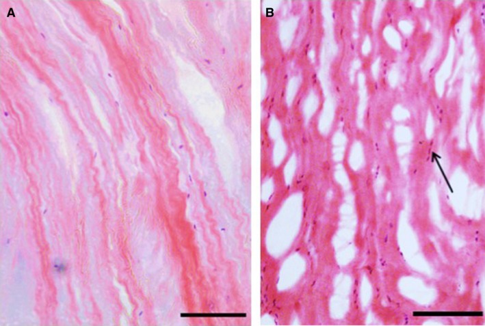
Outer annulus fibrosus (OAF) tissue stained with H&E. (A) Non‐degenerated scoliotic disc. Scale bar: 200 μm. (B) Degenerated spondylolisthesis disc. Scale bar: 100 μm. Note the elongated fibroblast‐like cells (arrows) aligned with the crimped collagen fibres, and the numerous microscopic tears and splits in (B).