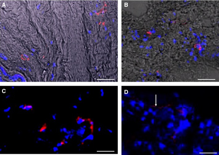
Figure 6.

Nerves in annulus tissues of herniated discs. Cell nuclei are stained blue with DAPI. Scale bar: 50 μm. (A) Fine nerves stained red for Substance P. Phase contrast imaging suggests matrix features. (B) Nerves stained red for PGP 9.5. (C) Nerves stained red for PGP 9.5. (D) Fine peripheral nerve (white arrow) stained red for Substance P.
