Figure 1.
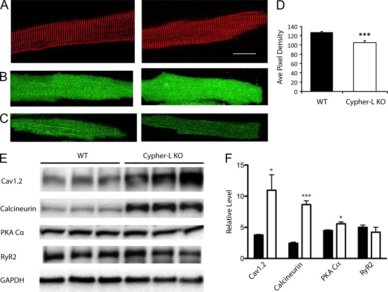
Morphology and protein expression in WT and LCyphKO mice. (A) Representative WT (left) and LCyphKO (right) dissociated cardiac ventricular myocytes immunostained for α-actinin to mark the z-lines. Bar, 25 µm. (B) Representative WT (left) and LCyphKO (right) dissociated cardiac ventricular myocytes immunostained for CaV1.2. (C) Confocal sections at the cell surface of representative immunostained WT (left) and LCyphKP (right) myocytes. (D) Quantification of CaV1.2 channels on the cell surface of ventricular myocytes from WT and LCyphKO (Cypher-L KO) mice. (E) Immunoblots of the indicated proteins in ventricular tissue from WT and LCyphKO mice. (F) Quantification of the indicated proteins in ventricular tissue from WT (black bars) and LCyphKO (white bars) mice (n = 4–6 replicates; 8–10 mice). *, P < 0.05; ***, P < 0.005. Error bars indicate SEM.

