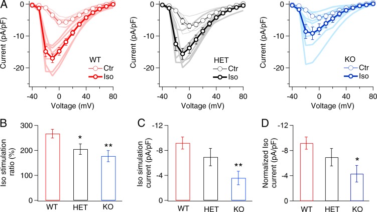
Figure 3.
β-adrenergic regulation of CaV1.2 Ca2+ current was reduced in myocytes from LCyphKO mice. (A) Mean peak current–voltage relations in the absence or presence of 100 nM Iso recorded in cardiac myocytes derived from WT, LCyphHET (HET), and LCyphKO (KO) mice. (B) Stimulation ratio for Ca2+ currents after treatment with 100 nM Iso in WT, LCyphHET, and LCyphKO mice, calculated as total current/basal current at a test potential of 0 mV. (C) Net β-adrenergic–stimulated Ca2+ currents induced by 100 nM Iso in WT, LCyphHET, and LCyphKO mice calculated as Iso current/basal current. (D) Normalized net β-adrenergic–stimulated Ca2+ currents induced by 100 nM Iso in WT, LCyphHET, and LCyphKO mice calculated as Iso current/basal current. WT, −9.2 ± 1.0 pA/pF; LCyphHET, −6.9 ± 1.4 pA/pF; LCyphKO, −4.3 ± 1.3 pA/pF; P < 0.05. n = 6–7 cells for each I-V curve; n = 3–4 mice for each I-V curve. *, P < 0.05; **, P < 0.01. Error bars indicate SEM.

