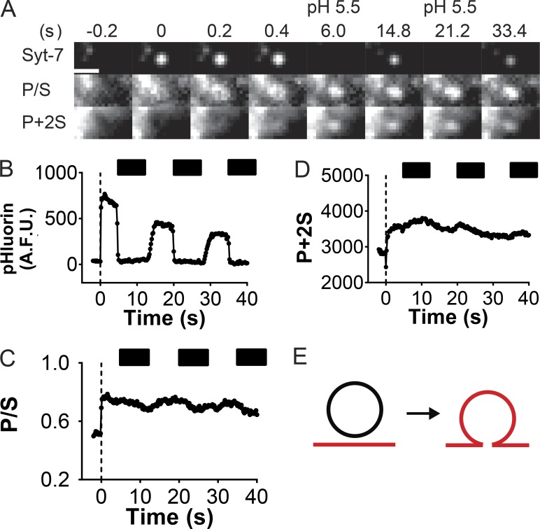
Figure 4.
Fusion pores of Syt-7–bearing dense-core vesicles are characterized by slow expansion. (A) Time series of a pHluorin-tagged Syt-7–bearing vesicle undergoing exocytosis with associated changes in DiD-labeled membrane fluorescence (P/S and P+2S). Bar, 960 nm. The cell was periodically perfused with a low-pH (5.5) solution to verify that the fusion pore was still open (note quenching of pHluorin fluorescence at times 6.0 and 21.2 s). (B–D) Intensity-versus-time curves for images in A. The dotted black line indicates the fusion frame (time 0). Black bars indicate pH 5.5 wash. A.F.U., arbitrary fluorescence units. (E) Simulations based on P/S and P+2S fluorescence emission suggest that the fusion pore is not expanding or is expanding slowly. Modified from Rao et al. (2014).

