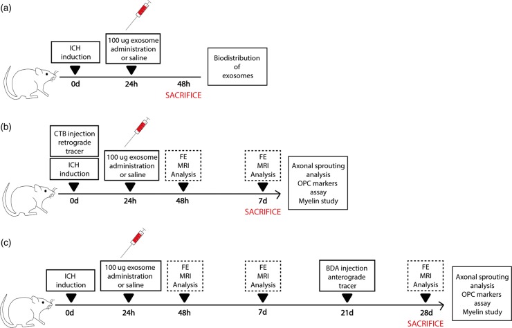
Figure 1.
Experimental protocol schematic. (a) Rats were subjected to an ICH by collagenase IV injection. Twenty-four hours later, rats received treatment (saline or exosomes). At 48 h, histological studies for biodistribution of exosomes were performed. (b) Rats were injected with collagenase IV to induce hemorrhagic stroke. During the same surgery, animals were inoculated with CTB as retrograde tracer. At 24 h poststroke, treatment (saline or exosomes) was administered through the tail vein. Later, 48 h after ICH and on day 7, behavior and imaging studies were evaluated. Seven days after ICH, histological studies were analyzed. (c) Rats were subjected to an ICH by collagenase IV and received treatment 24 h later (saline or exosomes). Forty-eight hours after ICH, on days 7 and 28, behavior and imaging studies were evaluated. BDA was injected as anterograde tracers 21 days after ICH. Histological studies were analyzed 28 days after ICH.

