Fig. 1.
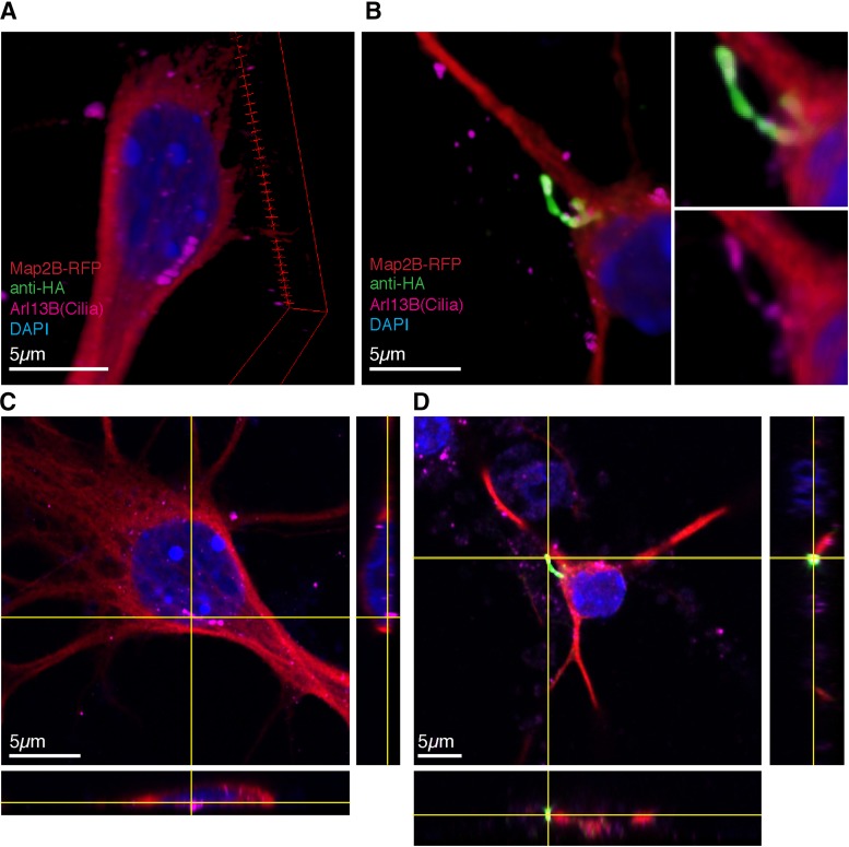
Rescued 5-HT6R localizes to the primary cilium in 5-HT6R-KO neurons. Super-resolution images taken on Zeiss LSM 880 with Airyscan of primary striatal/cortical primary neuronal cultures. Primary cultures from 5-HT6R-KO mice were transfected with 30% Map2B-RFP (Red) ± (A) empty vector or (B) 15% WT-HA-5-HT6 receptor plasmid, fixed and imaged. (C and D) XYZ projected images demonstrating colocalization of 5-HT6R (green) with Arl13B (cilia marker magenta) on the primary neuronal cilium.

