
Figure 3.
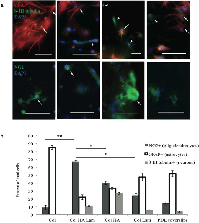
In vitro differentiation of rat embryonic spinal NPCs within Col HA Lam hydrogels resulted in the highest percent of oligodendrocyte differentiation.
(a) After five days in culture, cells were stained with anti-GFAP (red, top) to identify astrocytes, anti-β-III tubulin (green, top) to identify neurons, and anti-NG2 (green, bottom) to identify oligodendrocytes. DAPI (blue, top and bottom) was used to identify cell nuclei. All cell types were present in each of the four hydrogel compositions. However, the morphology and differentiation profiles were different in each gel type. The Col HA Lam hydrogel had the highest percentage of cells positive for oligodendrocyte markers compared to other hydrogel types. Scale bars = 50 μm. (b) E14-E15 rat spinal progenitor cells were cultured in hydrogels for 5 days in DMEM F12 medium supplemented with 1% fetal bovine serum. Note: n=10/group, this experiment was repeated twice. Note: ** p ≤ 0.001, * p ≤ 0.01