
Figure 5.
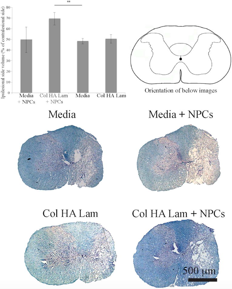
Larger spared tissue area around lesions in Col HA Lam hydrogel with NPCs group.
Two weeks post treatment, lesion extent was calculated by examining the difference in tissue volume between the ipsilesional and contralesional sides of the spinal cord at the epicenter of the lesion. Significantly more tissue was present at the epicenter in the Col HA Lam with NPCs transplantation group compared to media alone groups. Spinal cord cross-sections were stained with Nissl and Eriochrome cyanine (myelin stain). Note, scale bar is 500 μm, **p ≤ 0.01.