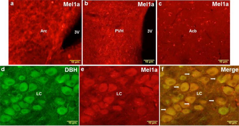
Fig. 3.

Representative higher magnification images of the Arc (a), PVH (b), Acb (c) and LC (d-f) showing single MEL1a (red), single DBH (green) and colocalized DBH + MEL1a (arrows) immunostaining. 3V, third ventricle; Acb, nucleus accumbens; Arc, arcuate nucleus; LC, locus coeruleus; PVH, paraventricular hypothalamic nucleus. n = 15. Scale bar = 10 μm.
