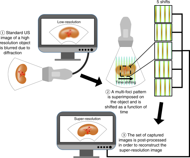
Fig. 1.
Schematic illustration of ASI steps of operation (left to right). An object that contains sub-diffraction features is imaged with ultrasound. A multifocal pattern is generated at the position of the object and the echoes are captured by the transducer. Five emitted fields, corresponding to five shifts of the pattern, are transmitted sequentially, and a set of five images is captured. The set of captured images is post processed to reconstruct the super-resolution image, where sub-diffraction features are visible in comparison to the original low-resolution image

