Fig. 8.
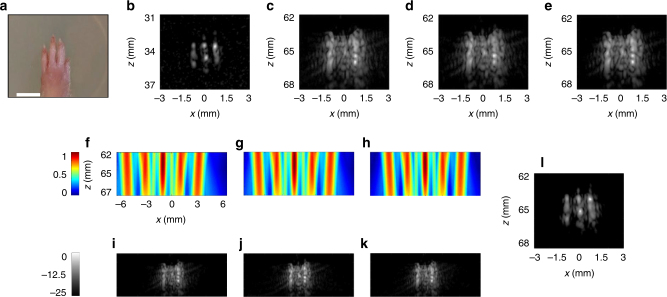
Experimental results obtained by imaging an ex vivo paw from a Sprague–Dawley rat. Results are presented in 25 dB dynamic range log scale with jet and gray colorbars common to all subfigures. Axes are common to f–k. a An image of the sample. The ultrasound images were acquired along the phalanges distal to 4th phalange. White scale bar represents 3 mm. b High-resolution image captured at z = 34 mm, where the phalanges are resolvable. For all other images, the position of the paw was shifted to 65 mm. c Plane wave image. d The result of coherently compounding plane wave images acquired from five angles. e Two-way focused image. f–h Three predicted emitted pressure fields. i–k Corresponding images. l Super-resolution reconstructed image. Example selected from n = 3 repetitions

