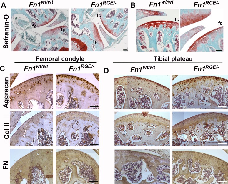
Fig 1. Morphology of articular cartilage in Fn1RGE/- mice.
A) Representative Safranin-O stained sections from wild type (Fn1wt/wt) and mutant (Fn1RGE/-) knee joint cartilage from 5-month-old male mice. Femoral condyle (fc) and tibial plateau (tp) are shown. B) High magnification showing the femoral cartilage. The dotted line indicates the tidemark that separates the non-calcified (superficial) from the calcified zone. C, D) Hematoxylin and immunoperoxidase staining to detect aggrecan, collagen type II and FN in the femoral condyle (C) and tibial (D) articular cartilage. Scale bars, 50 μm.

