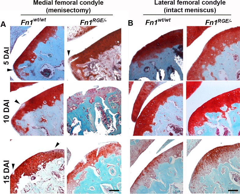
Fig 3. OA progression in Fn1wt/wt and Fn1RGE/- cartilage exposed to high mechanical load.
A) Safranin-O staining of representative medial femoral knee cartilage sections from control and Fn1RGE/- mice after exposure to high load at 5, 10 and 15 days after OA induction (DAI). B) Controls of the contralateral femoral condyles stained with Safranin-O. Scale bars, 50 μm.

