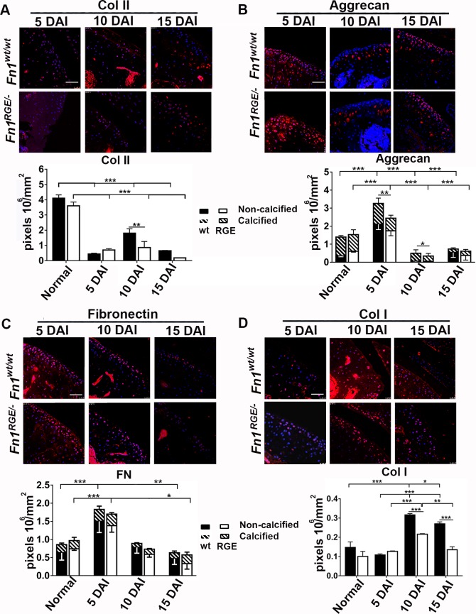
Fig 4. Immunofluorescence and quantification of ECM components in the femoral knee cartilage after exposure to high load.
A) Immunofluorescence of collagen type II and quantification of pixel density per mm2 in the articular cartilage zone. Staining of cartilage from untrained (normal) mice is shown in Fig 2C. Aggrecan (B), FN (C) and collagen type I (D) staining and pixel density in articular cartilage. The pixel densities of aggrecan and FN were determined in the non-calcified and calcified zones. Results represent mean ± SEM. Scale bars, 50 μm. Statistical significances: *p<0.05, **p<0.01 and ***p<0.001.

