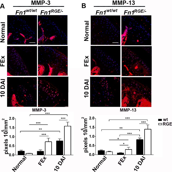
Fig 5. Immunofluorescence and quantification of MMP-3 and MMP-13 in femoral knee cartilage exposed to high load.
A) Immunofluorescence staining for MMP-3 levels and quantification of pixel density per mm2 in the articular cartilage of mice 10 days after exposure to normal activity (normal), moderate load (FEx) and high load (10 DAI). B) MMP-13 levels. Values represent mean ± SEM. Scale bars, 50 μm. Statistical significances: *p<0.05, **p<0.01 and ***p<0.001.

