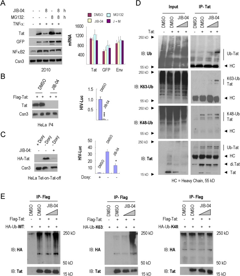
Fig 6. JIB-04 increases Tat K63Ub and proteolytic destruction.
(A) Left, immunoblot analysis of HIV-1 Tat expression in 2D10 cells exposed to DMSO, JIB-04, MG132 or JIB-04+MG132. Cells were pre-treated with or without TNFα (10 ng/ml) for 16 h. The COP9 signalosome complex subunit 3 (Csn3) served as loading control. Right, qRT-PCR analysis of Tat, GFP and Env mRNA levels in these cells. Values shown in the Y-axis were normalized to mRNAs from 2D10 cells without TNFα-stimulation. (B) Dual-Luc (HIV-LTR-Luc/SV40-Renilla-Luc) reporter gene analysis in FLAG-Tat101 transfected HeLa P4 cells. Left, immunoblot analysis of FLAG-Tat101 protein levels in cells treated with DMSO or 2 μM JIB-04. Right panels show dual-luc reporter gene activity in these cells. Significant differences between HIV-Luc activity treated by DMSO or 2 μM JIB-04 were calculated by Student’s T-test (*p<0.05, **p<0.005, ***p<0.0005). (C) Dual-Luc reporter gene analysis, as in part B, in Tet-on-Tat-off HeLa cells. Left, immunoblot analysis of HA-Tat86 protein levels in 2D10 cells treated by DMSO or 2.5 μM JIB-04. Right, dual-Luc reporter gene activity in these cells. Significant differences between HIV-Luc activity treated by DMSO or 2.5 μM JIB-04 were calculated by Student’s T-test (*p<0.05, **p<0.005, ***p<0.0005). (D) Analysis of the effect of JIB-04 on endogenous Tat K63Ub levels in HeLa cells. HIV-1 Tat was immunoprecipitated from lysates of HeLa cells exposed to DMSO or JIB-04 (1 μM and 3 μM), and endogenous ubiquitylation was monitored using the antisera indicated to the left of each panel (HC = antibody heavy chain). (E) Immunoprecipitation of FLAG-Tat-101 from lysates of HeLa cells treated with DMSO or JIB-04 (1μM and 3μM). Ubiquitination of FLAG-Tat-101 in the presence of ectopically expressed HA-ubiquitin-WT, HA-ubiquitin-K63-only or HA-ubiquitin-K48-only was assessed using anti-HA antisera.

