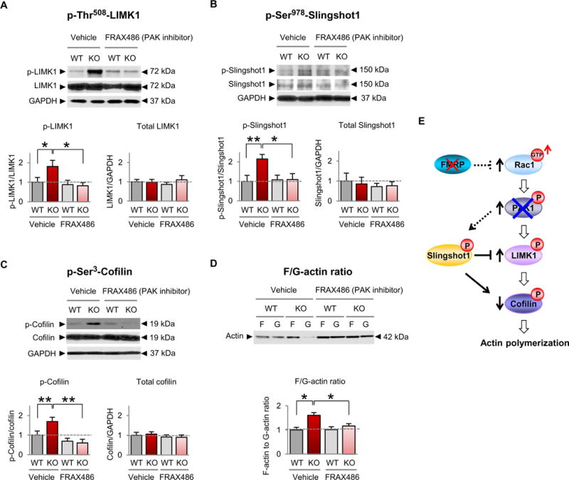
Fig. 3. Inhibition of PAK restores cofilin signaling and actin polymerization in FXS model mice.

(A to D) Representative Western blots and summary data for p-Thr508-LIMK (A), p-Ser9780-Slinghot1 (B), p-Ser3-cofilin (C), and the F-actin/G-actin ratio (D) in somatosensory synaptosomes from 1-week-old male WT and Fmr1 KO mice given a single subcutaneous injection of FRAX486 or vehicle [20% (w/v) hydroxypropyl-β-cyclodextrin] 8 hours before sacrifice (WT vehicle, n ≥ 12; KO vehicle, n ≥ 10;WT FRAX486, n ≥ 8;KO FRAX486, n ≥ 9). We note that overall there was no significant difference in total actin abundance in synaptosomes from FRAX486- versus vehicle-treated mice. Data are means ± SEM. *P < 0.05, **P < 0.01. (E) Schematic of Rac-cofilin signaling depicting proposed mechanism by which inhibition of group 1 PAKs (PAK1, PAK2, and PAK3) restores the abundance of p-LIMK1, p-Slingshot1, p-cofilin, and actin polymerization in Fmr1 KO animals.
