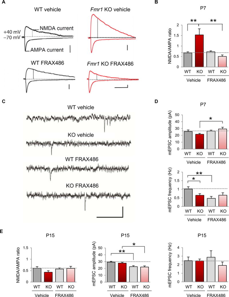
Fig. 5. Inhibition of PAK corrects functional synaptic deficits in layer V of the somatosensory cortex.

(A) Representative evoked EPSC recordings from male WT (left) and Fmr1 KO (right) mice, either vehicle-treated (top) or FRAX486-treated (bottom) at P7. AMPAR-mediated EPSCs were measured as the peak current at −70 mV, and the NMDA component was measured by depolarizing the cell to +40 mV and measuring the current 60 ms after the onset of the outward current in the presence of 50 μM picrotoxin. Calibration: 100 ms, 50 pA. (B) Summary data of the NMDA/AMPA ratio in all recordings (WT vehicle, n = 13; Fmr1 KO vehicle, n = 11; WT FRAX486, n = 11; Fmr1 KO FRAX486, n = 6). (C) Representative traces of mEPSC recordings from male WT and Fmr1 KO animals, vehicle- or FRAX486-treated at P7. Calibration: 500 ms, 50 pA. (D) Summary data of mEPSC amplitude and frequency in all recordings (WT vehicle, n = 10; Fmr1 KO vehicle, n = 10; WT FRAX486, n = 11; Fmr1 KO FRAX486, n = 9). (E) Summary data for all recordings at P15 (NMDA/AMPA ratio: WT vehicle, n = 15; Fmr1 KO vehicle, n = 7; WT FRAX486, n =11; Fmr1 KO FRAX486, n = 10; mEPSC amplitude and frequency: WT vehicle, n = 18; Fmr1 KO, n = 7; WT FRAX486, n = 16; Fmr1 KO FRAX486, n = 12). Data are means ± SEM. *P < 0.05, **P < 0.01.
