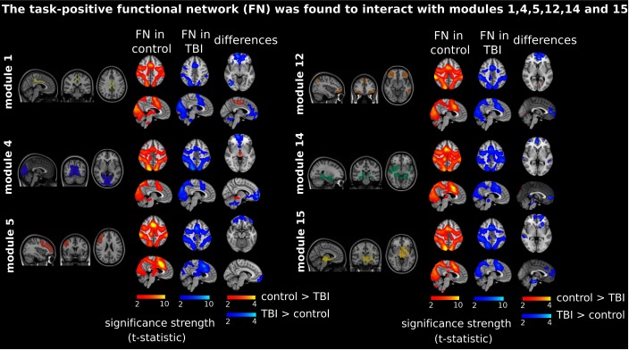
Figure 4. .
Prefrontal recruitment into the task-positive functional network (FN). These are depicted as in Figure 3, but the most representative cluster now resembles the task-positive network (see the labels “FN in control” and “FN in TBI”), which is now resulting from Module 1 (posterior cingulate cortex), Module 4 (medial visual cortex), Module 5 (medial frontal gyrus), Module 12 (inferior parietal and temporal gyrus, lateral frontal orbital gyrus, rostral pars of middle frontal gyrus, and pars orbitalis and triangularis), and Modules 14 and 15 (subcortical structures). Similar to what is shown in Figure 3, now TBI patients recruited the prefrontal part of the brain in interaction with the task-positive network (colored in blue in the “differences” column).

