. 2018 May 1;7:e35731. doi: 10.7554/eLife.35731

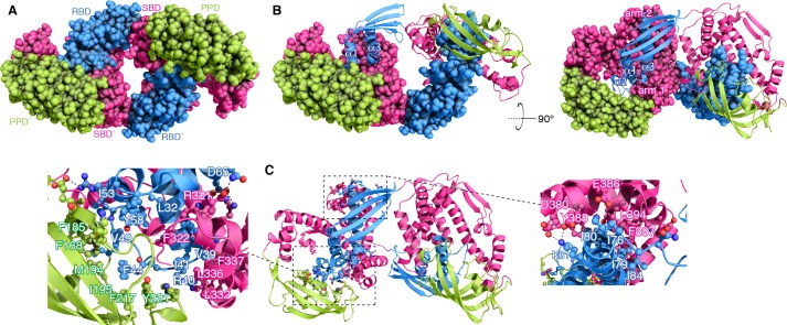
Figure 2. Structural basis for TF dimerization.

(A) The lowest-energy structure of the TF dimer is shown as space-filling model. TF forms a dimer in a head-to-tail orientation. RBD, SBD, and PPD are shown in blue, magenta, and green, respectively. (B) One of the TF subunits is shown as space-filling model and the other subunit shown in ribbon. The helices of the RBD and the two arm regions are labeled. (C) Expanded views of the dimeric interfaces highlighting contacts between the two subunits. Residues involved in mediating dimerization are shown as ball-and-stick.

Figure 2.

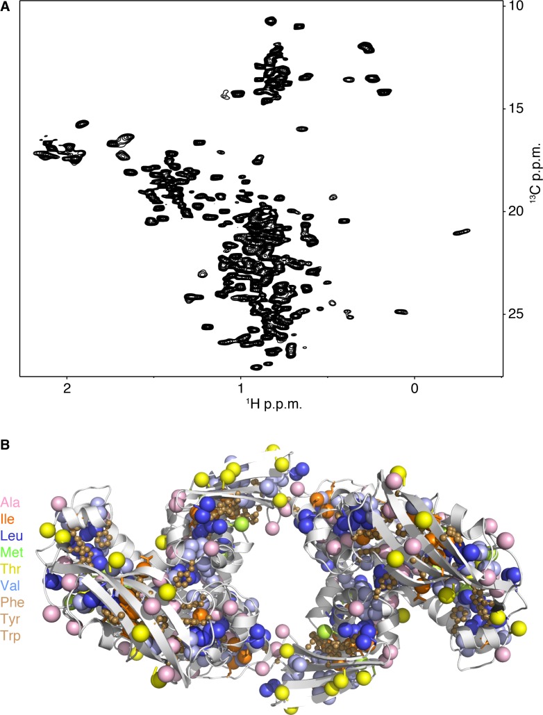
Figure 2—figure supplement 1. NMR of dimeric TF.

Figure 2—figure supplement 1.

(A) 1H–13C methyl HMQC spectrum of [U-2H; Ala-13CH3; Met-13CH3; Ile-δ1-13CH3; Leu,Val-13CH3/13CH3; Thr-13CH3]-labelled TF. (B) TF is enriched in hydrophobic amino acids, such as methyl-bearing (Ala, Ile, Leu, Met, Thr and Val) and aromatic (Phe, Tyr and Trp).
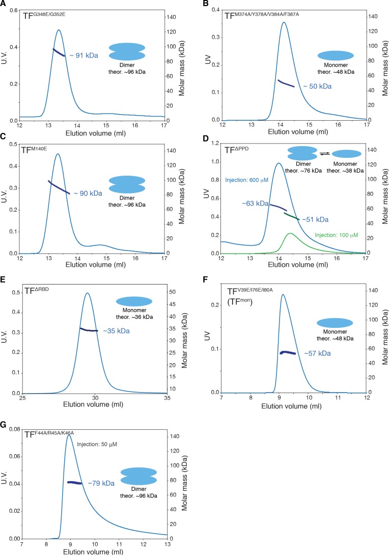
Figure 2—figure supplement 2. SEC-MALS of TF mutants.

Figure 2—figure supplement 2.

SEC-MALS profiles of TFG348E/G352E (A), TFM374A/Y378A/V384A/F387A (B), TFM140E (C), TFΔPPD (D), TFΔRBD (E), TFV39E/I76E/I80A (TFmon) (F), and TFF44A/R45A/K46A (G) are shown. Proteins were injected at the concentration of 100 μM unless otherwise stated. TFG348E/G352E, TFM374A/Y378A/V384A/F387A, and TFM140E have mutations on the substrate-binding site A, B, and D, respectively. TFF44A/R45A/K46A has mutation at the ribosome-binding loop containing the signature motif. Destabilization of the dimer by the introduction of mutations on the substrate-binding site B as well as by deletion of PPD containing site E supports the engagement of these substrate-binding sites in the dimerization (Figure 3B). Moderate effect of the deletion of PPD to dimerization implies that the contribution of the interaction between PPD and RBD in the formation of the dimer is less significant and is auxiliary. The mutations in the signature motif have little effect to the dimer formation, which is consistent with the fact that only a part of the ribosome-binding loop is involved in the dimer formation and the rest is floating in the cavity formed by PPD and SBD (Figure 3—figure supplement 1A and B).
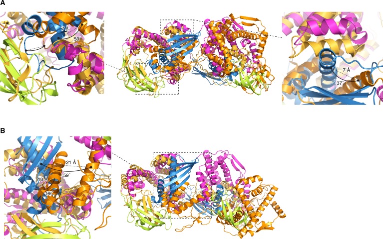
Figure 2—figure supplement 3. Comparison with PRE-based docking models.

Figure 2—figure supplement 3.

The structure of TF dimer superimposed with previously reported PRE-based docking models (Morgado et al., 2017); conformer 1 [PDB code: 5OWI] (A), and conformer 2 [PDB code: 5OWJ] (B). PPD, SBD, and RBD in the structure of TF dimer are shown in green, pink, and blue, respectively. The docking model is shown in orange and gold. The coordinates are superimposed on the backbone heavy atoms of SBD. Differences in rotation and translation of the helices in RBD between the previously reported PRE-based docking model and the current dimer structure are indicated. Most of the contacts, which are seen in the current structure of the TF dimer and were validated by mutagenesis and chemical shift perturbation mapping, are not present in the PRE-based models. In the PRE-derived conformer 1, RBD makes contacts with the arm 1 and the PPD of the other subunit, which were also seen in the current dimer TF structure. However, in the PRE-derived conformer 1 the RBD makes no significant contacts with the arm 2, which is not consistent with the significant effect that mutations in the arm 2 have on the dimerization as shown by SEC-MALS (Figure 2—figure supplement 2B). The PRE-derived conformer 2 has RBD snagged on the tips of the arm 1 and arm 2, as well as on the edge of PPD. A very small overlap between the substrate-binding sites and the dimer interface is seen in the PRE-derived conformer 2. In contrast, in the current structure of TF dimer, RBD is buried inside the cradle formed by SBD and PPD of the other subunit, thus explaining why the TF dimer dissociates upon binding to the substrate protein (Figure 1—figure supplement 1A,E and F). In addition to the significant differences in the overall domain orientation between the PRE-derived models and the current TF structure, the RBD structure itself appears to be loosely packed in the PRE-derived models.
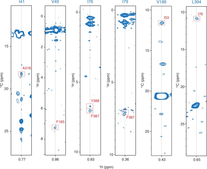
Figure 2—figure supplement 4. Examples of the inter-molecular NOEs.

Figure 2—figure supplement 4.

Representative strips from 13C-edited NOESY-HMQC and HMQC-NOESY-HMQC NMR experiments. The intermolecular NOE cross peaks are designated by a dashed-line red rectangle.