. 2018 May 1;7:e35731. doi: 10.7554/eLife.35731

Figure 3. Conformational changes of TF upon dimerization.

(A) The structure of one subunit in the TF dimer (colored as in Figure 1A) and the crystal structure of monomeric TF (colored grey) [Protein Data Bank (PDB) code: 1W26] are superimposed for SBD. The changes in rotation and translation of the RBD and PPD between the monomer and the dimer are indicated. (B) View of the structure of dimeric TF highlighting the positioning of the substrate-binding sites (colored orange). The five main substrate-binding sites are labeled A, B, C, D, and E.

Figure 3.

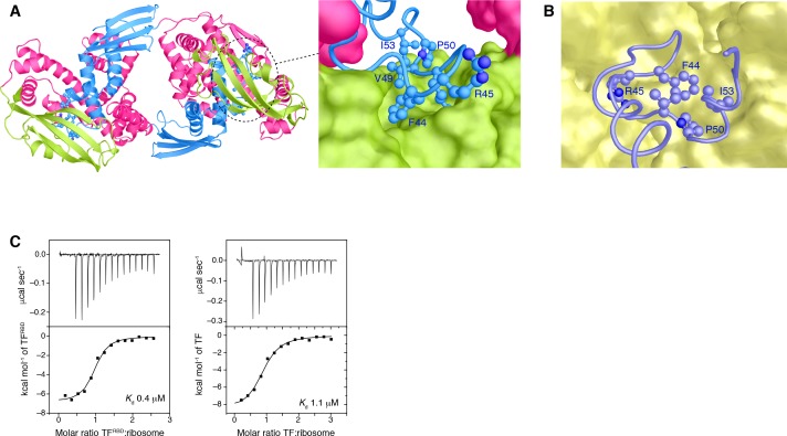
Figure 3—figure supplement 1. The ribosome-binding loop in the TF dimer.

Figure 3—figure supplement 1.

Close-up view of the ribosome-binding loop in the TF dimer (A) or TF in complex with the ribosome (PDB ID: 1W2B) (B). The amino acid residues of the ribosome-binding loop involved in the interaction are shown in ball-and-stick. PPD, SBD, and RBD are shown in green, pink, and blue, respectively. The ribosome is represented as yellow surface model. (C) ITC traces of the titration of TF (right) and RBD (left) to the ribosome. Titration of RBD indicated slightly stronger affinity than that of TF, which is consistent with the fact that RBD is responsible for the binding to the ribosome and the ribosome-binding loop is protected in the TF dimer. The experiments were performed at 22°C.
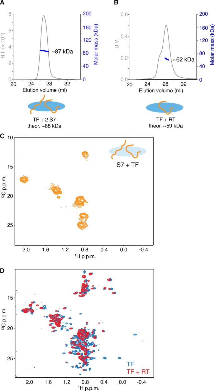
Figure 3—figure supplement 2. Characterization of small substrate proteins in complex with TF.

Figure 3—figure supplement 2.

(A) SEC-MALS of E. coli S7 in complex with TF indicating two S7 molecules bind to the monomer of TF. (B) SEC-MALS of E. coli reverse transcriptase (RT)-Ec86 255–320 in complex with TF indicating one RT molecule binds to the monomer of TF. Unliganded dimeric TF eluted before TF-RT complex, resulting in a shoulder on the left side of the main peak. (C) 1H-13C methyl HMQC of [U-2H; Met-13CH3; Ala-13CH3; Ile-δ1-13CH3; Leu,Val-13CH3/13CH3]-labeled S7 in complex with unlabeled TF showing narrow dispersion of the resonances of methyl groups in S7. (D) Overlay of 1H-13C methyl HMQC of [U-2H; Met-13CH3; Ala-13CH3; Ile-δ1-13CH3; Leu,Val-13CH3/13CH3]-labeled TF (blue) and TF-RT complex (red). Several resonances that broaden out in complex with RT are located at the substrate-binding sites on TF. Most of the dispersed resonances observed in the spectrum of TF-RT complex match to the resonances of TF and no dispersed resonances are found for RT, which suggests that RT in complex with TF does not form a folded structure. The results indicate that both of RT and S7 binds to monomeric TF as an unfolded state, although the possibility of the existence of minor folded population cannot be excluded.