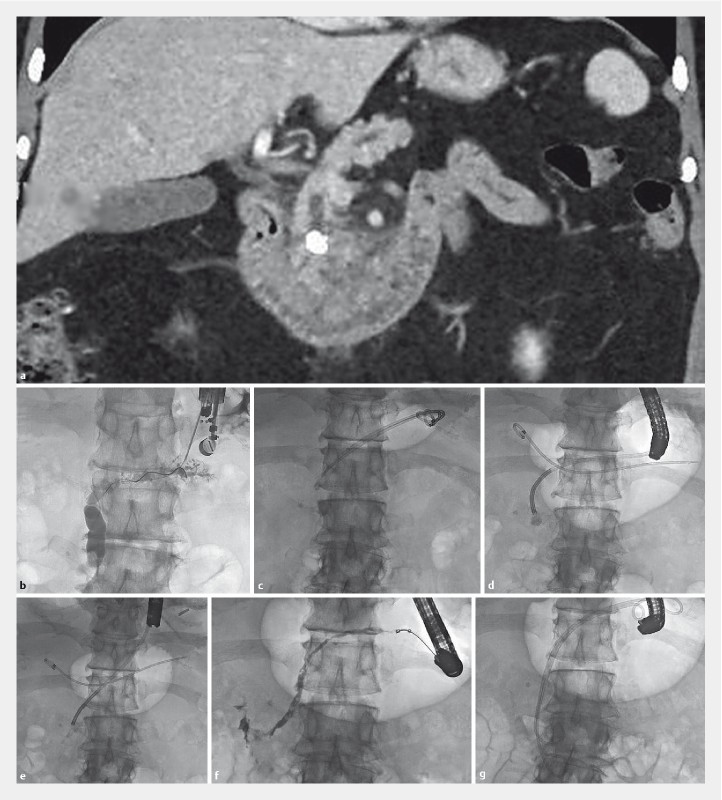
Fig. 1 a.

Coronal CT scan image showing intraductal obstructing pancreatic head stone. b Radiographic image during EUS-guided pancreaticogastrostomy. Guidewire is advanced through a 19G needle into the duct. c Radiographic scout image at follow-up ERCP showing PG stents within the duct. d Radiographic image of pancreatoscope dvanced to the level of the stone. Note one of the prior PG stents is free in the stomach overlying the image. e Radiographic image after stone fragmentation, f Follow-up antegrade pancreatogram showing free flow into the duodenum. g Radiographic image showing two double pigtail transgastric/transpapillary 7Fr stents placed.
