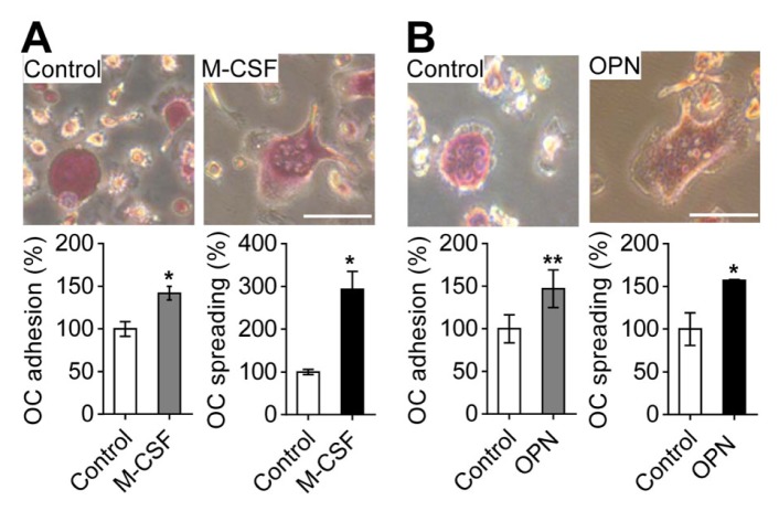
Fig. 1.
M-CSF- and OPN-induced osteoclast adhesion and spreading. (A) Effects of M-CSF on osteoclast adhesion and spreading. For osteoclast adhesion and spreading induced by M-CSF, suspended osteoclasts treated with M-CSF (30 ng/ml) were seeded on culture plates and incubated for 1 h (for cell adhesion) or 4 h (for cell spreading). (B) Effects of OPN on osteoclast adhesion and spreading. For osteoclast adhesion and spreading by OPN, osteoclasts were incubated in OPN-coated culture plates for 1 h (for cell adhesion) or 4 h (for cell spreading). The attached osteoclasts were fixed and stained with TRAP. Osteoclast adhesion and spreading were assessed by counting the number of TRAP-positive multinucleated osteoclasts and by measuring the surface area of osteoclasts, respectively. The upper panel shows representative phase-contrast images. Data are expressed as the mean ± standard deviation (S.D.) (n = 3) and presented as mean percentage relative to control. *P < 0.01; **P < 0.05. Scale bars, 50 μm.

