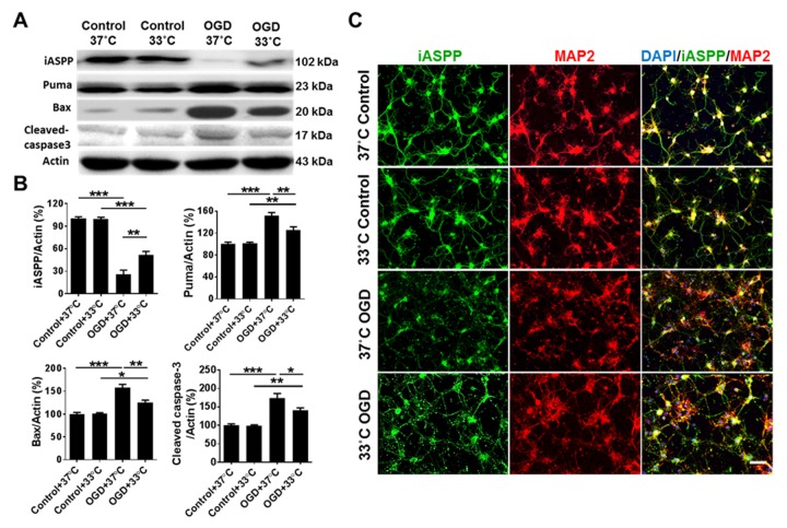
Figure 1.
Mild therapeutic hypothermia regulates the expression of iASPP and its targets in primary neurons treated with OGD/R. (A) Representative protein in bands of iASPP, Puma, Bax and cleaved caspase-3 at 24 h after OGD from the Western blot. Actin served as a loading control. (B) Densitometric quantification of iASPP, Puma, Bax and cleaved caspase-3 from the Western blot, n = 4 per group. (C) Representative images showing expression of iASPP (green) in primary neurons (red) treated with OGD at 37°C or 33°C. DAPI was used as a nuclear marker. Bar, 50 μm.*p < 0.05, **p < 0.01, ***p < 0.001, by one-way ANOVA and Tukey’s test.

