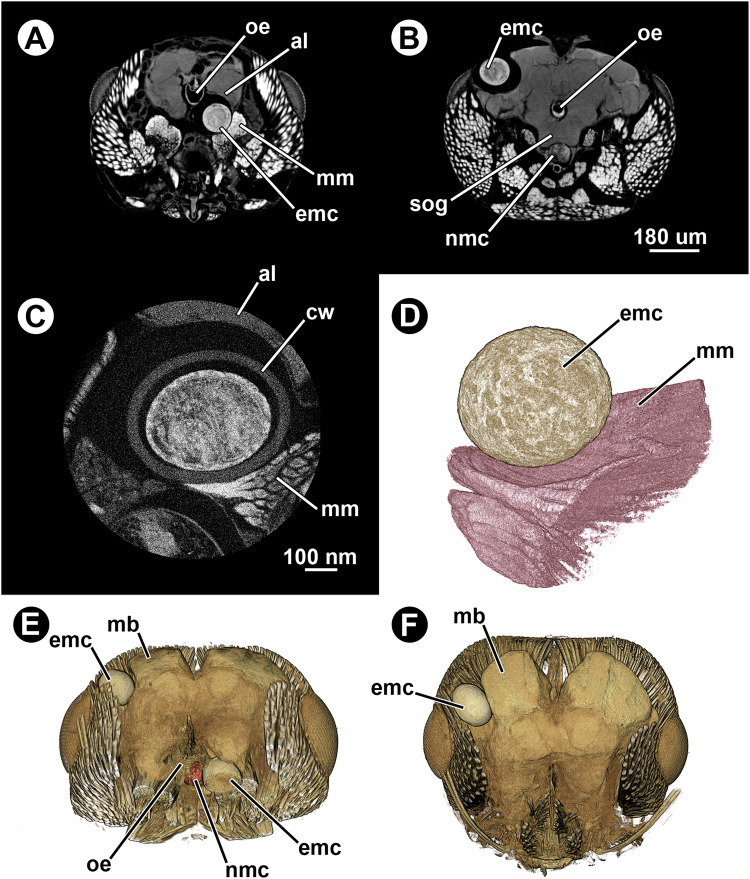
Figure 6.
Micro-CT-based images of an infected ant head (Formica aserva) harbouring two encysted metacercariae. (A), (B) and (C), virtual cross sections. (D) False-coloured 3D volume rendering of one of the encysted metacercariae and the surrounding musculature. (E) False-coloured 3D volume rendering of the ant head harbouring two encysted metacercariae; cross section in dorsofrontal view. (F) False-coloured 3D volume rendering of the ant head harbouring two encysted metacercariae; horizontal view. Abbreviations: al, antennal lobe; cw, cyst wall; emc, encysted metacercaria; mb, mushroom bodies; mm, mandibular muscle; nmc, non-encysted metacercaria; oe, oesophagus; sog, suboesophageal ganglion.

