Fig. 1.
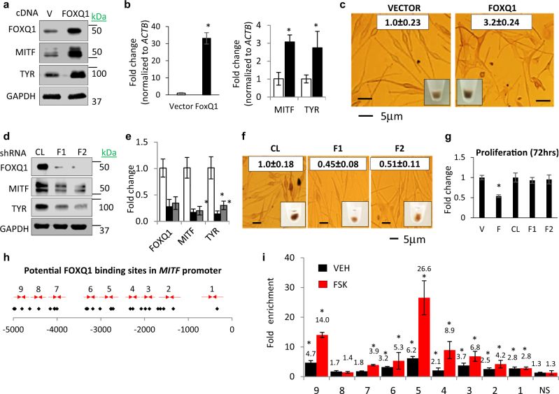
FOXQ1 induces MITF-dependent differentiation. a, b NHM transduced with empty vector (VECTOR) or FOXQ1-expressing vector (FOXQ1) were probed in immunoblotting with indicated antibodies (left) or in Q-RT-PCR (right). FOXQ1/ACTB, MITF/ACTB, and TYR/ACTB signal ratios are shown. c Cells described in a were imaged as adherent cells or as cell pellets followed by quantification of total melanin content (white boxes). d, e Cells transduced with control (CL) or FOXQ1 (F1, F2) shRNAs were probed in immunoblotting with the indicated antibodies (left panel) or in Q-RT-PCR (right panel). FOXQ1/ACTB, MITF/ACTB, and TYR/ACTB signal ratios are shown. f NHM described in d, e) were imaged as adherent cells or as cell pellets followed by quantification of total melanin content (white boxes). g NHM expressing empty vector (V), FOXQ1 cDNA f, or control (CL) or FOXQ1 shRNAs (F1, F2) were counted for 72 h starting 48 h post infection. The cell numbers at 72 h were normalized by those before plating and by the ratio of these numbers in vector or control cells. h Human MITF promoter. Shown are FOXQ1-binding sites (diamonds) and PCR primers (arrows). i Q-PCR signals in reactions with DNA precipitated with FOXQ1-specific antibodies from untreated and FSK-treated NHM cells were normalized by corresponding signals in DNA precipitated with IgG antibodies and by signals obtained with MITF nonspecific control primers (NS). All data represent mean ± SEM. Statistical significance was assessed using two-tailed Student’s t-tests. A p < 0.05 (*) was considered significant

