
Figure 4.
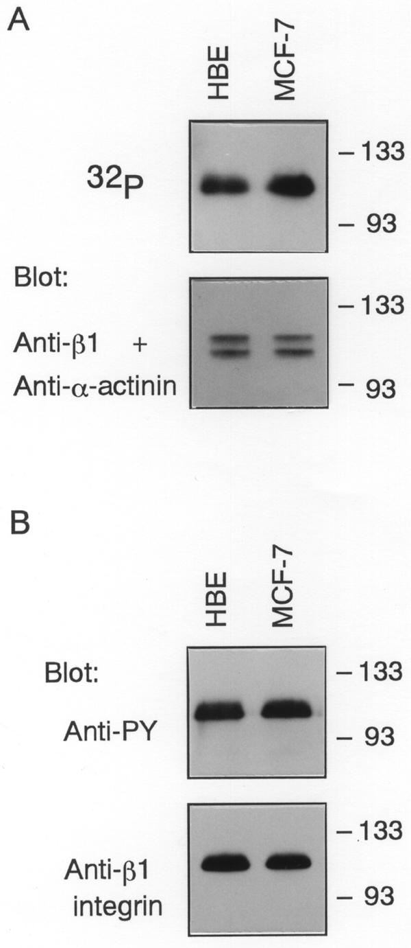
The phosphorylation state of β1 integrin. (A) HBE or MCF-7 cells which were quiescent and adherent to collagen IV were metabolically labeled with [32P]orthophosphoric acid and β1 integrin was immunoprecipitated from the cells. After separation on 8% SDS-PAGE, gels were dried and autoradiographed. Unlabeled samples were electrophoresed and transferred onto membranes. The blots were probed with antibodies to β1 integrin and α-actinin. Molecular size markers are indicated at right in kDa. (B) Tyrosine phosphorylation of β1 integrin in HBE and MCF-7 cells. Integrin β1 was immunoprecipitated from HBE or MCF-7 cells and the immunoprecipitates were resolved by 8% SDS-PAGE before blotting. The blots were then probed with anti-PY or anti-integrin β1 monoclonal antibody. Molecular size markers are indicated at right in kDa.