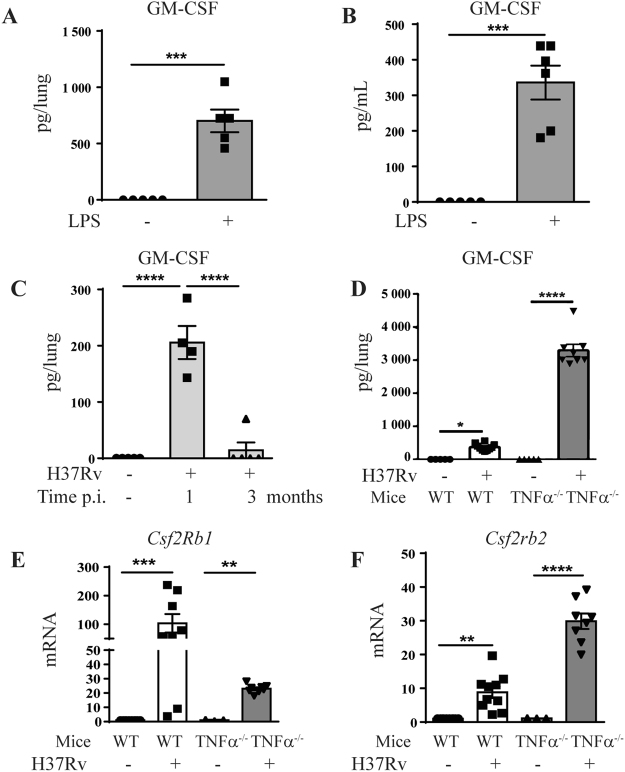
Figure 1.
Increased pulmonary GM-CSF after acute airway inflammation and M. tuberculosis infection in vivo. WT mice were challenged intranasally with saline or LPS (1 µg/mouse) and GM-CSF levels in lung (A) and bronchoalveolar lavage (BAL) fluid (B) were measured after 24 h by ELISA. Pulmonary concentration of GM-CSF protein was measured 1 and 3 months (C) after in vivo M. tuberculosis infection (1000 ± 200 CFU/mouse i.n.) in WT mice, of after 1 month in WT and TNFα−/− mice (D). Data are representative of two independent experiments and are expressed as mean ± SEM (n = 5–8 mice per group). The expression of GM-CSF receptor β subunits genes Csf2rb1 (E) and of Csfrb2 (F) in the lungs 4 weeks post-M. tuberculosis infection in WT and in TNFα−/− mice was analyzed by microarray50. Each group of infected mice has been statistically compared to the uninfected mice of the same phenotype. ****p < 0.0001, ***p < 0.001, **p < 0.01, *p < 0.05.

