Fig. 2.
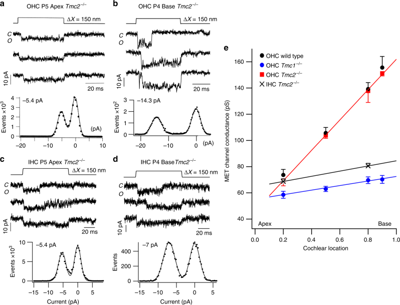
Tonotopic organization of MET channels. a Three examples of single-channel currents in response to hair bundle deflection (top) in P5 apical Tmc2−/− mice. Amplitude histogram of events gives −5.4 pA single-channel current. b Three examples of single-channel currents in P4 basal OHC; amplitude histogram of events gives −14.3 pA single-channel current. Holding potential −84 mV. c Three examples of single-channel currents evoked in apical IHC of P5 Tmc2−/− mice. Amplitude histogram of events gives 5.4 pA single-channel current. d Three examples of single-channel currents in P4 basal IHC; amplitude histogram of events gives 6.9 pA single-channel current. Holding potential −84 mV. e Collected results on MET channel conductance plotted against fractional distance along the cochlea from the apex, for OHCs of P3–P7 wild-type (black symbols), Tmc2−/− mice (red symbols), and Tmc1−/− mice (blue symbols) and for Tmc2−/− mouse IHCs (black crosses). Each point is the mean ± SEM; numbers of cells, apex to base: OHC wild type, 14, 4,14, 7; OHC Tmc2−/− 11, 4, 10, 3; OHC Tmc1−/−18, 3, 14, 3; IHC Tmc2−/− 7, 5. Lines drawn through points for Tmc2−/− and Tmc1−/− mice. MET channels for Tmc2−/− mice still exhibit a tonotopic conductance gradient like wild type, but the gradient is virtually absent in Tmc1−/− mice indicating the tonotopic conductance gradient is supported by TMC1 but not TMC2. All measurements in 1.5 mM extracellular Ca2+

