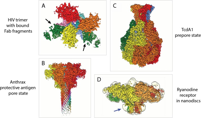
Fig. 4.
Selected examples of biomedically relevant cryo-EM structures. (A) Cryo-EM structure of the trimeric envelope glycoprotein of HIV. It was solved in complex with two neutralizing antibody Fab fragments. [EMD-3308]. (B) Cryo-EM structure of the anthrax protective antigen pore from Bacillus anthracis. [EMD-6224]. (C) Cryo-EM structure of TcdA1 from Photorhabdus luminescens in its prepore state. [EMD-3645]. (D) Cryo-EM structure of the ryanodine receptor RyR1. Map at low threshold (transparent) is shown to visualize the nanodisc (blue arrow), which stabilizes the transmembrane helices of RyR1. [EMD-2751]. Individual subunits are depicted in various colors. The hetero-trimeric HIV envelope glycoprotein in A can be divided into the gp120 trimer (yellow, orange, and red) and gp41 trimer (pink, blue, and cyan). Bound Fab fragments (green) are indicated by black arrows. Heptameric anthrax protective antigen pore (yellow, orange, light red, dark red, magenta, cyan, and green) in (B), pentameric TcdA1 (yellow, orange, red, green and blue) in (C), and tetrameric RyR1 (yellow, orange, red, and green) in D

