
Figure 2.
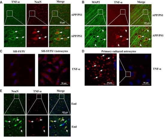
Localization of TNF-α in the brains of AD patients and APP/PS1 Tg mice. (A,B) The brains of 3-month-old APP/PS1 Tg mice were collected after anesthesia and perfusion (n = 10). The slices of mouse brains were double-stained with NeuN (red) and TNF-α (green) (A) or MAP2 (green) and TNF-α (red) antibody (B) before observation using confocal microscopy. (C) The astrocytes were co-cultured with neurons using transwell experiments for 24 h. The neurons were then immunostained with TNF-α antibody before observation under confocal microscopy. (D) Primary cultured astrocytes were immunostained with a TNF-α antibody before observation under confocal microscopy. (E) The tissue blocks of human brains at a late stage of AD were collected from the New York Brain Bank at Columbia University. Forty micrometer free-floating slices were prepared using a cryostat (n = 1). The slices of human brains were double-stained with NeuN (green) or TNF-α (red) antibody before observation using confocal microscopy.