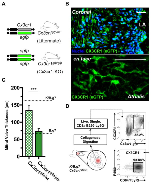
Figure 2. K/B.g7 cardiac valve inflammation and fibrosis requires CX3CR1.
A, Cx3cr1-gfp mice contain an eGFP reporter construct in the endogenous Cx3cr1 locus; mice homozygous for the eGFP allele are Cx3cr1-null (Cx3cr1-KO) whereas heterozygous mice retain Cx3cr1 expression. B, Top: Cx3cr1-eGFP+ cells are seen throughout the MV interstitium of K/B.g7:Cx3cr1gfp/wt mice (coronal sections, nuclear counter-staining with Hoechst 33342); bottom: Cx3cr1-eGFP+ cells in inflamed K/B.g7 MVs exhibit a characteristic phagocyte morphology (whole-mount MVs from K/B.g7:Cx3cr1gfp/wt mice, processed using tissue clearing methods and imaged en face). C, MV thickness measurements from Cx3cr1-KO animals (Cx3cr1gfp/gfp) relative Cx3cr1-replete controls (Cx3cr1gfp/wt) (median MV thickness at 8 weeks: 132.9 μm [n=4] and 69.1 μm [n=10], Cx3cr1gfp/wt and Cx3cr1gfp/gfp, respectively); reference thicknesses for MVs from K/B.g7 and B.g7 control mice are provided. D, Flow cytometry on collagenase-2-DNAse-I-digested MVs from K/B.g7:Cx3cr1gfp/wt mice demonstrating GFP+ MNPs (CD45.2+CD3e−B220/CD45R−Ly6G−CX3CR1+) uniformly display a phenotype consistent with macrophages (CD64/FcγRI+), therein. Scale bars in B are equal to 50 microns. Asterisks (***) in C indicate statistically significant differences at p<0.005.

